Large Scale SVD for 3-D Reconstruction of Images from 2-D Projections
Through Analysis of Projections from Electron Micrographs
The project involves the identification and classification of
2-dimensional electron micrograph images of biological macromolecules
and the subsequent generation of the corresponding high resolution 3-D
images. The underlying algorithm is based upon the statistical
technique of principle component analysis. An SVD of the data set is
performed to extract the largest singular vectors which are then used
in the classification procedure.
Example
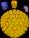
 Description: 26 Å map of HSV-1 A capsid reconstructed from 140
particles selected from 400-kV spot-scan electron micrographs of the
A-capsids of HSV-1 embedded in vitreous ice. A-capsid has a triangulation
number of 16. Each of the 60 asymmetric units consists of one penton
subunit, one P-hexon, one C-hexon, half an E-hexon, and six types of
connecting triplexes (Ta, Tb. Tc, Td, Te, and 1/3 Tf). The top inserts
show a computationally isolated hexon (blue), penton (yellow) and
triplex (pink).